Muscle is one of the most fundamental of animal tissues. It is muscles that animate our bodies — allowing us to move, stand upright, breathe, and even speak. Muscles are precisely the sort of thing that we might expect a designer of embodied intelligent beings to produce. At the very least, on the supposition of intelligent design, the existence of muscles is not particularly surprising. But how surprising is the existence of muscles under the perspective of naturalism?
The number of muscles in the human body exceeds six hundred. The majority are attached by tendons to the skeleton — their primary purpose being to move the skeleton. When skeletal muscles contract, they shorten and pull the bone. The contraction of muscles also generates heat, which contributes to the maintenance of our core body temperature. The muscular system is unable to perform its job of animating the skeleton without assistance from the nervous, respiratory, and circulatory systems. The electrochemical impulses that drive muscle contraction are transmitted by motor neurons (the nervous system). Muscle cells contain many mitochondria, which perform cellular respiration, generating the ATP needed for contraction. This depends critically on the exchange of the gases oxygen and carbon dioxide between the blood and air by means of the respiratory system. The oxygen is brought to the muscles, and carbon dioxide removed, by means of the circulatory system. Thus, a tremendous amount of functional integration is required for muscle contraction to work. You can find a short video animation and summary of the structure of skeletal muscle in this video.
In a series of two articles, I will summarize the incredible design of muscles that allows them to fulfil their function. In the first part, we shall review the structure and arrangements of muscles, as well as muscle tone and sense. In a second article, I shall explain the fascinating biochemistry of muscle contraction. I hope that the reader will get a sense for how improbable all this is from the vantage of the naturalistic hypothesis. The information that follows is well established and can be found in any decent textbook on anatomy and physiology.
The Structure of Muscles
Each skeletal muscle contains many thousands of cells called muscle fibers, or myocytes. The number of muscle fibers that contract depends on the task being performed. For example, picking up a book requires the contraction of more finger-flexing muscle fibers than picking up a pencil, which requires the contraction of only a small number of fibers.
The muscle fibers are organized into bundles called fascicles. These fascicles are given structural support by a connective tissue that surrounds them called the perimysium. The perimysium also protects and distributes the blood vessels and nerves that supply the muscle, in addition to facilitating the transmission of forces generated by muscle contractions. Together with the endomysium (connective tissue that surrounds individual muscle fibers) and the epimysium (connective tissue that surrounds the entire muscle), the perimysium helps to maintain the overall integrity and organization of the skeletal muscle. The structure of skeletal muscles is shown in the figure below:
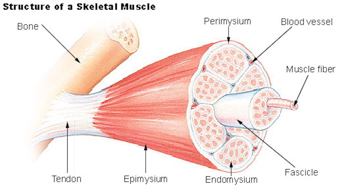
Image source: Wikimedia Commons.
Having a muscular, nervous and respiratory system will do one no good, however, unless the muscles are attached to the skeleton. Muscles are attached to bones by tendons, which are tough, fibrous, connective tissues. When a muscle contracts, it generates force, which is transmitted to the bones by tendons, allowing the limb or body part to move. Tendons also provide stability to joints — acting as stabilizing elements that prevent excessive or unwanted movement, helping to maintain joint integrity during muscular contractions. Generally, a muscle possesses at least two tendons, each of them connected to a different bone. The attachment point where the muscle’s tendon connects to a less movable bone is referred to as the origin, whereas the attachment point where the muscle’s tendon connects to a more movable bone is called the insertion. When the muscle contracts, it pulls the insertion bone towards the origin, resulting in joint movement.
The Arrangement of Muscles
There are two common types of arrangements of muscles across joints and around the skeleton — namely, opposing antagonists and cooperative synergists. Antagonistic muscles are pairs of muscles that have opposite actions at a joint. One muscle in the pair is responsible for producing a specific movement, while the other has an opposing action. For example, the biceps and triceps in the upper arm are a classic example of antagonistic muscles (see the figure below).
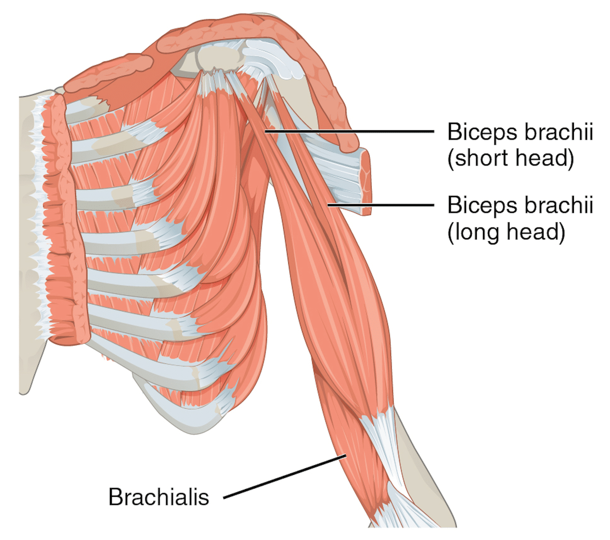
Image source: CFCF, CC BY-SA 4.0 https://creativecommons.org/licenses/by-sa/4.0, via Wikimedia Commons.
When you flex your elbow, the biceps (located on the front of the upper arm) contract to bend the arm, while the triceps relax and lengthen. Muscles are unable to push, exerting no force when they relax. Thus, the elbow can be flexed by the biceps but cannot be extended, and thus we need another muscle called the triceps (located on the back of the upper arm). When you extend your elbow, the triceps contract and the biceps relax.
Synergistic muscles, by contrast, work together to produce the same movement at a joint. They assist the prime mover in performing a specific action, helping to stabilize the joint and provide additional force or control to the movement. For example, when you flex your elbow, the biceps are the prime mover, but other muscles (the brachialis and brachioradialis) act as synergists to assist the biceps in generating the movement. These synergistic muscles provide support and help fine-tune the motion. You might wonder why we need three muscles to carry out the same task. When your hand is positioned palm up, the prime mover (which does most of the work of flexing) is the biceps. When your hand is positioned palm down, the prime mover is the brachialis. When your hand is positioned thumb up, the prime mover is the brachioradialis. Thus, depending on the position of your forearm, different muscles can be more or less effective at generating force.
Synergists can also serve to steady or stabilize a joint, rendering it possible to make more precise movements. For example, when drinking a glass of water, the prime mover for flexing the arm is the biceps. To assist in getting the water to your mouth and not spilling it down your chin or over your shoulder, the joint is stabilized by the shoulder muscles.
The Role of the Brain in Muscle Movement
The region of the brain responsible for generating the nerve impulses for movement is the frontal lobes of the cerebrum (the frontal lobes are labelled in the figure given below).
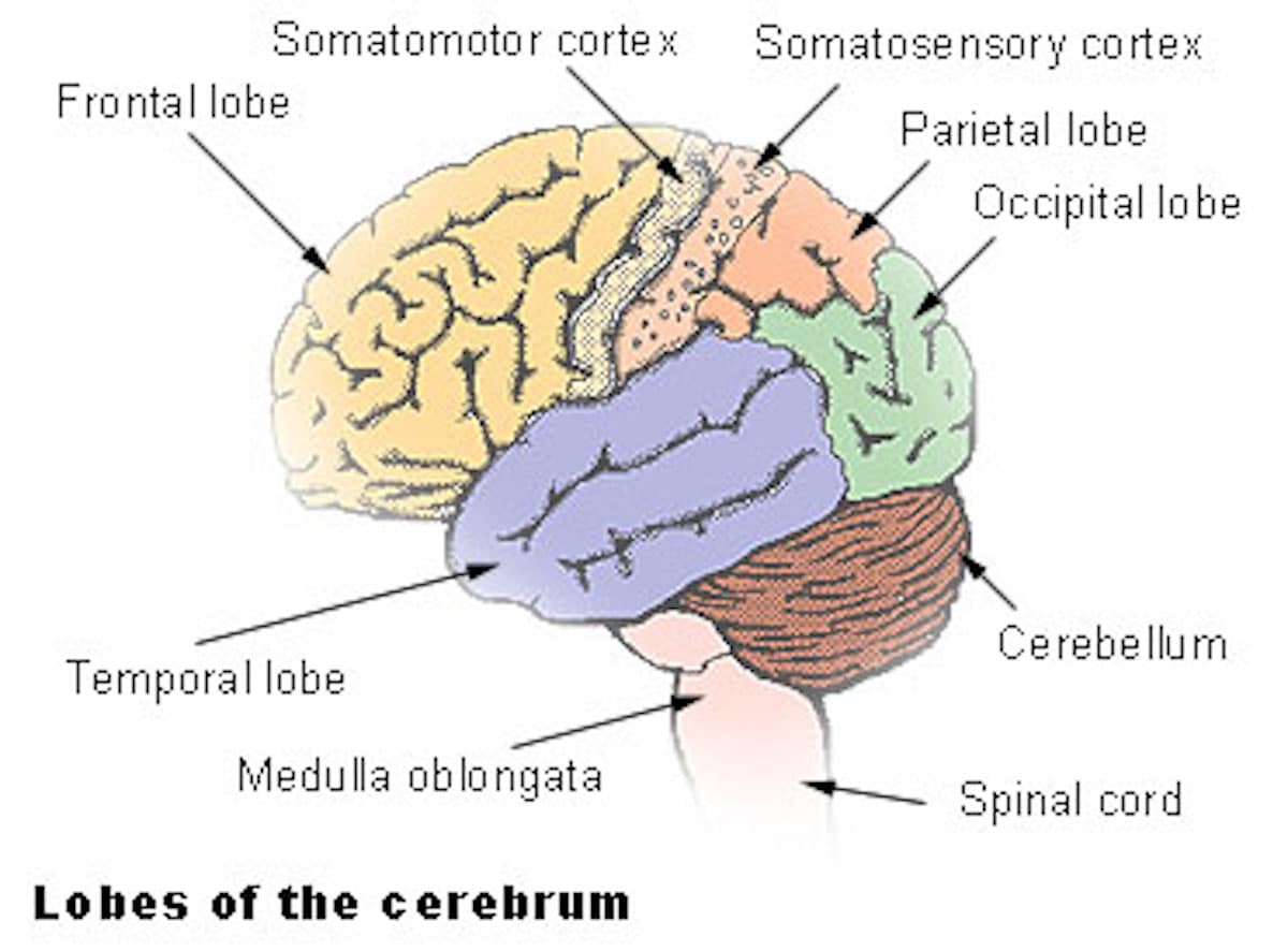
Image source: Wikimedia Commons.
The muscle fibers contract when they receive electrochemical impulses generated from the motor areas of the frontal lobes, that travel along motor neurons (grouped into nerves). A single neuron can innervate anywhere between a few to hundreds of muscle fibers, as its axon can branch extensively (this is referred to as a motor unit).
In muscles that carry out small and precise movements (such as those responsible for moving the eyes or fingers), muscles typically have small motor units (2 to 100 muscle fibers per neuron). On the other hand, muscles that have to carry out powerful rather than precise movements (for example, the large muscles of the hips and legs) have hundreds of muscle fibers per neuron.
The cerebellum (also labelled in the figure above) is the part of the brain responsible for regulating coordination and motor control, operating largely below the level of consciousness. This means that many of its functions occur without our awareness. The cerebellum receives input from multiple sensory systems, including the proprioceptive system (information about the body’s position and movements), the vestibular system (balance and spatial orientation), and the visual and auditory systems. These inputs help the cerebellum establish a sense of where the body is in space and how it is moving. The cerebellum also receives information from the motor cortex, which provides “efference copies” of the motor commands sent to muscles. Efference copies are predictions of the intended motor output and are used to compare with the actual sensory feedback. This comparison helps the cerebellum detect any discrepancies between the intended and actual movements. The cerebellum acts as an integration center for sensory and motor information. It constantly compares the efference copies with the incoming sensory feedback, such as proprioceptive signals from stretch receptors and Golgi tendon organs, as well as visual and vestibular input. This comparison occurs at the subconscious level, allowing the cerebellum to detect errors in movement even before they become apparent to the conscious mind.
When the cerebellum detects errors in movement or discrepancies between the intended and actual outcomes, it generates corrective signals. These signals are sent to the motor cortex and other motor control centers in the brain. The cerebellum adjusts the ongoing motor commands to correct the errors and improve the precision and accuracy of movements.
The cerebellum is also involved in motor learning and adaptation. Through repetitive practice and learning, the cerebellum stores information about various motor tasks and their associated sensory feedback. This allows it to refine movements over time, even without conscious awareness. For example, when you learn to ride a bike, the cerebellum helps you automatically adjust your balance and coordination without needing to consciously think about it.
The cerebellum also plays a role in feedforward control, where it predicts the sensory consequences of planned movements. It can make anticipatory adjustments to movements based on the expected sensory feedback. For example, when you reach for an object, the cerebellum can adjust the motor commands to account for the expected weight and resistance of the object, allowing for smoother and more precise movements.
The cerebellum also receives information from inner ear receptors for equilibrium and uses it to balance the contractions of antagonistic muscles such that the contractions of one set do not cause the body to fall over.
Muscle Tone
With the exception of certain stages of sleep, the majority of our muscles exist in a state of slight contraction, called muscle tone. This enables us to keep an upright posture. Only a few muscle fibers in the muscle have to contract in order for the muscle to be in a slightly contracted state. To prevent the muscle from becoming fatigued, alternate fibers take turns at contracting. This is subconsciously regulated by the cerebellum. The heat produced by muscle fibers during cellular respiration (necessary for production of ATP) accounts for roughly 25 percent of the total body heat at rest.
Muscle Sense
Muscle sense (also known as proprioception) is the body’s ability to sense and perceive the position, movement, and tension of one’s muscles and joints. It plays a crucial role in maintaining balance and coordinating movement. It operates in the background of our conscious awareness, enabling us to perform various tasks without having to constantly think about the position of our limbs or the force required for specific actions. Activities that involve fine motor skills (such as typing on a keyboard or playing on a musical instrument) depend heavily on muscle sense for control and accuracy, and these improve with repetition and practice due to what is called muscle memory. As neural pathways that control the necessary movements become strengthened, the experienced pianist or typist need not consciously think about every movement.
Muscles contain receptors known as stretch receptors (otherwise known as muscle spindles or proprioceptors), which detect changes in muscle length when it is stretched. The brain interprets these sensory impulses to generate a mental image of where the muscle is in space. The impulses responsible for muscle sense are received and processed by in the cerebellum (for unconscious muscle sense) and the parietal lobes of the cerebrum (for conscious muscle sense).
Evidence of Design
As we have seen, multiple interdependent systems are required for the muscular system to work — among those needed are the circulatory, respiratory, and nervous systems, in addition to the tendons that attach muscle to bone — not to mention the incredible structure and arrangement of the muscles themselves (containing many mitochondria to meet the energy demand; being comprised of thousands of muscle fibers; being arranged antagonistically and synergistically for coordinated action, etc). The origin of the skeletal muscles, then, depends on many co-dependent changes in order to come about. This is not particularly surprising in light of a design perspective, but becomes wildly surprising if we suppose the falsity of design. Thus, muscles provide powerful evidence of intelligent design.
In a second article, I shall review the process of muscle contraction, which (as we will see) takes the inference to design to an entirely new level.
Note: This article was originally published on October 26 2023 at Evolution News & Science Today.
1 thought on “The Incredible Design of Muscles”
Pingback: Understanding the Biochemistry — and Intelligent Design — of Muscle Contraction - Jonathan McLatchie | Writer, Speaker, Scholar
Comments are closed.