The ear is responsible for two of our most fundamental senses — hearing and equilibrium — the receptors for which are all found inside the inner ear. For an incredible animation of how hearing works, I recommend this YouTube video.
Here, I will describe the anatomy of the ear and the biological basis of the sense of hearing. The information below can be found in any good anatomy and physiology textbook. You can also find a good discussion of this subject in Chapter 11 of Your Designed Body, by Steve Laufmann and Howard Glicksman.
It might be helpful to refer to the illustration of the ear below as you read the description that follows.
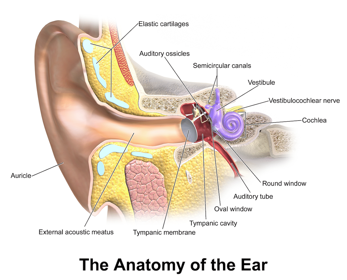
Image credit: Blausen.com staff (2014). “Medical gallery of Blausen Medical 2014”. WikiJournal of Medicine 1 (2). DOI:10.15347/wjm/2014.010. ISSN 2002-4436., CC BY 3.0 https://creativecommons.org/licenses/by/3.0, via Wikimedia Commons.
The Outer Ear
The outer ear is comprised of the auricle and ear canal. The auricle is composed of skin-covered cartilage. In dogs (who have movable ears), the auricle can serve as a funnel for sound waves. In humans, on the other hand, its absence would not negatively affect our hearing.
Skin containing ceruminous glands lines the ear canal (also called the external auditory meatus). The ear canal is a tube-like structure that extends from the outer ear to the middle ear. It is responsible for directing sound waves into the ear, which then travel through the ear canal and arrive at the eardrum (tympanic membrane) in the middle ear. The eardrum vibrates in response to these sound waves, and these vibrations are transmitted to the middle ear bones.
The Middle Ear
The middle ear is a cavity in the temporal bone that is filled with air. The tympanic membrane (popularly called the eardrum) is a thin, flexible membrane that separates the outer ear from the middle ear, and is stretched across the end of the ear canal. When sound waves enter the ear canal, they strike the eardrum, causing it to vibrate. Behind the eardrum, there are three small bones known as the ossicles — namely, the malleus (hammer), incus (anvil), and stapes (stirrup). The ossicles form a chain and are connected to each other. When the eardrum vibrates in response to waves, it causes the malleus to move, which, in turn, moves the incus and stapes. This mechanical linkage helps amplify the vibrations and transmits them from the eardrum to the inner ear.
The middle ear is also connected to the nasopharynx (back of the throat) through a tube called the eustachian tube. This tube helps to equalize air pressure on both sides of the eardrum. This is essential to maintain equilibrium of air pressure between the middle ear and external atmospheric pressure, to allow the eardrum to properly vibrate.
The Inner Ear
The inner ear is also a cavity within the temporal bone, and is also called the bony labyrinth. It is lined with a membrane called the membranous labyrinth. Between the bone and membrane is a fluid called perilymph, and within the membranous structures of the inner ear is a fluid called endolymph. Three of these structures (the utricle, saccule, and semicircular canals) are concerned with equilibrium. The other (the cochlea) relates to hearing.
The cochlea’s appearance is like the shell of a snail. The inside of the cochlea is partitioned into three canals, filled with fluid. The uppermost canal is called the scala vestibuli, and it is filled with perilymph (a fluid similar to cerebrospinal fluid). Sound vibrations travel through the cochlea and arrive at the scala tympani. The middle canal is called the scala media (otherwise known as the cochlear duct), and it is separated from the scala vestibuli by Reissner’s membrane, and from the scala tympani by the basilar membrane. The scala media contains endolymph and is where the sensory cells of the cochlea (known as hair cells) are located. These hair cells are of course not in fact hair, but, rather, are specialized microvilli that are responsible for converting sound vibrations into electrical signals that can be interpreted by the brain. Situated above the hair cells is the tectorial membrane which, as we shall see, is crucial for hearing.
The Sense of Hearing
The process of hearing begins with the production of sound waves, which are pressure fluctuations that are propagated through air. These waves are funneled into the ear canal by the pinna (the external part of the ear). The ear canal carries the sound waves to the eardrum (tympanic membrane), which causes it to vibrate. These vibrations are then transmitted to the malleus, incus, and stapes, which amplify the vibrations. The stapes is connected to the oval window, a membrane-covered opening to the inner ear. Vibration of the stapes bone against the oval window creates pressure waves in the fluid-filled cochlea. As the pressure waves pass through the fluid in the cochlea, they cause vibration of the basilar membrane. This results in the bending of hair cells against the tectorial membrane, which in turn triggers the release of neurotransmitters that convert mechanical vibrations into electrical signals. These electrical signals are transmitted to the brain by the auditory nerve, where they are interpreted as sound by the auditory areas in the temporal lobes of the cerebral cortex.
The auditory nerve fibers carrying information from one ear partially cross to the opposite side at a structure in the brainstem, known as the trapezoid body. This means that signals from both ears are sent to both sides of the brain. This plays an important role in sound localization and spatial processing, allowing the brain to compare the timing and intensity of signals from both ears, helping us to determine the direction of a source of sound. Impulses arriving from each inner ear are counted and compared by the auditory areas, to determine the direction of a sound. If there are more impulses coming from the right cochlea than from the left one, the brain projects the sound to the right, and vice versa.
The neurons of the auditory cortex are organized in a manner similar to a piano keyboard — being arranged from low to high pitch. The brain is also able to detect volume, rhythm, and tempo, as well as timbre, which is a quality of tone (a guitar playing a middle C and a piano playing the same note at the same volume will sound different due to the unique timbre of each instrument).
Masterpiece of Engineering
The anatomy of hearing described above is of course the system found in humans and other terrestrial mammals. Many other organisms have less advanced hearing systems. For example, fish lack external ears and have structures called otoliths that detect vibrations and changes in water pressure. Reptiles, birds, and amphibians also often lack an external ear and have a single middle ear bone instead of the three found in mammals. And most invertebrates (such as crustaceans and mollusks) lack ears and a sense of hearing altogether. Typically, claims that the sense of hearing evolved by natural selection focus on these as intermediate stages. The incus, malleus, and stapes are thought to have arisen from three reptilian bones associated with the jaw — the quadrate bone, articular bone, and columella respectively.
However, the vertebrate sense of hearing involves several fundamental anatomical features that are common to all vertebrate hearing systems and cannot be removed without severely compromising (or completely eliminating) the ability to hear. For example, the cochlea (which contains the hair cells) is a critical component for transducing sound vibrations into electrical signals that the brain can interpret. Indeed, the leading cause of hearing loss is damage to the hair cells. Furthermore, the auditory nerve, which carries electrical signals from the hair cells in the cochlea to the brain, is crucial for transmitting auditory information to the central nervous system. In injuries or infections (such as meningitis) where the auditory nerve is damaged, the result can be a complete and permanent loss of hearing in that ear. The eardrum (tympanic membrane), which vibrates in response to sound waves, transmitting these vibrations to the middle ear ossicles, is also an essential aspect of the sense of hearing. If the eardrum is damaged or perforated, the consequence can be deafness. A minimum of one middle ear ossicle appears to be essential for hearing as well. Another crucial feature of the auditory system is the oval window, the membrane-covered opening between the middle and inner ear, located at the base of the stapes bone. Vibrations transmitted by the ossicles are transferred to the fluid within the cochlea through the oval window.
Thus, several different structural components are necessary for the vertebrate sense of hearing. It strains credulity to suppose that an unguided process of random variation sifted by natural selection could assemble such a delicately arranged system. It instead points to a cause with foresight.
Note: This article was originally published on November 30 2023, at Evolution News & Science Today.