In a previous article, I discussed the irreducible complexity of sperm cells and the seminal fluid for successful fertilization. Now, I will review the exquisite design features of a female egg cell (also called an ovum, plural ova). Here is an animation of the incredible process of reproduction, from ejaculation to birth.
Oogenesis
Oogenesis (the process of egg cell formation) begins during embryonic development when the primordial germ cells are specified. These cells migrate to the genital ridges, which later develop into the female ovaries. Prior to birth, the primordial germ cells undergo mitotic divisions to form oogonia, the precursor cells for eggs. These oogonia transform into primary oocytes, which are diploid cells arrested in prophase I of meiosis. This arrest typically occurs before or shortly following birth.
Primary oocytes are surrounded by somatic cells to form primordial follicles, which go through a process called folliculogenesis, where they develop into primary, secondary and eventually tertiary follicles. As a female reaches sexual maturity, some primary oocytes are activated each menstrual cycle. The activated primary oocyte completes meiotic division I, resulting in the formation of a secondary oocyte and a smaller cell called a polar body (the primary purpose of the polar body is to discard the extra genetic material that is produced during meiosis). However, the secondary oocyte is arrested in metaphase II.
The mature follicle ruptures during ovulation, releasing the secondary oocyte into the fallopian tube. If fertilized by a sperm cell, the secondary oocyte completes meiotic division II, resulting in a mature egg (ovum) and another polar body. If fertilization does not occur, meiosis II is not completed. After ovulation, the remaining follicle transforms into the corpus luteum, which secretes hormones like progesterone to prepare the uterus for a potential pregnancy. If fertilization doesn’t happen, the corpus luteum degenerates, resulting in a drop in hormone levels. This triggers menstruation, and the cycle resets.
Fertilization
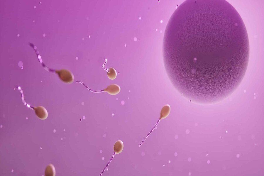
As I discussed previously, sperm cells swim through the female reproductive tract, directed by the cilia, in addition to chemical signals. Chemicals called chemoattractants are released by the egg cell, and these serve as signaling molecules that generate a concentration gradient. The sperm cell is capable of chemotaxis, a process that results in the sperm cell moving up the concentration gradient, towards higher chemoattractant concentrations. Changes in chemoattractant concentration are detected by specialized receptors on the surface of sperm cells. When an increase in concentration is detected, a signaling cascade is triggered within the cell, which influences the flagellum’s beating pattern. Thus, the sperm moves progressively in the direction of the egg — that is, the source of the chemoattractants. As the sperm swims towards the egg, the concentration of chemoattractants is continuously being measured, which allows it to adjust its course in order to fine-tune its movements. Once the sperm gets within close proximity of the egg, it encounters other signaling molecules that further guide the sperm cells and direct it towards the egg’s plasma membrane, the site of fertilization.
Upon reaching the egg, the sperm cell encounters the zona pellucida, a glycoprotein rich matrix that surrounds the egg. Sperm-egg recognition begins with the interaction between glycoproteins on both the sperm surface and zona pellucida, thereby guiding the sperm cell towards the egg cell’s surface.
In a previous article, I wrote about the acrosome, a specialized structure possessed by sperm cells, that contains enzymes that aid in penetrating the egg’s protective barriers. Contact between the sperm and the zona pellucida results in the acrosome undergoing exocytosis, releasing these enzymes. These enzymes help to create a pathway for the sperm to arrive at the plasma membrane of the egg. Once through, fusion occurs between the egg and the sperm’s plasma membrane, thereby allowing the sperm’s genetic material to come into proximity with the egg’s cytoplasm.
Egg Activation
Upon fusion of the plasma membranes of the sperm and egg, various changes are triggered in the egg, collectively referred to as “egg activation.” First, the egg’s membrane becomes less permeable to other sperm, in order to prevent a single egg from being fertilized by more than one sperm cell. The fast block to polyspermy involves a change in the electrical properties of the egg’s plasma membrane. When the sperm’s outer layers are successfully penetrated by the sperm cell, it triggers the release of calcium ions (Ca2+) from intracellular stores in the egg.
The influx of calcium ions serves as a signal to initiate changes in the egg’s membrane potential. Ion channels on the egg’s membrane are opened, and facilitate the entry of sodium ions (Na+). The consequence is that the egg’s plasma membrane depolarizes. Normally, the egg’s membrane is maintained at a negative resting potential. However, the influx of positive sodium ions neutralizes this negative potential, making the membrane potential less negative. The altered membrane potential makes it more difficult for other sperm to initiate the fusion process, and thereby creates a temporary electrical barrier that inhibits additional sperm from fusing with the egg. The depolarization is a temporary phenomenon. After a brief period, the egg membrane potential is restored to its normal resting state (often referred to as “resetting” the egg).
A secondary defense against polyspermy is known as the slow block, or the “cortical reaction.” As calcium ions are released upon fertilization, this triggers the exocytosis of cortical granules, located just beneath the egg’s plasma membrane, containing enzymes. The glycoproteins in the zona pellucida are cross-linked by these enzymes, and this results in the hardening of the zona pellucida, reducing its permeability. The modified zona pellucida forms a structure called the “fertilization envelope,” which surrounds the egg, forming a barrier that physically blocks additional sperm from gaining access to the egg’s surface.
Changes also take place in the egg cell that promote the completion of meiosis and initiate early embryonic development. The genetic material of the sperm and egg, consisting of a single set of chromosomes each (23 chromosomes in humans), combine to form a diploid cell called the zygote, which contains the full set of chromosomes needed to develop a new individual. This instantly determines gender, eye and hair color, and many other traits.
After fertilization has occurred, the zygote begins to undergo a series of rapid cell divisions through a process called cleavage. This results in the development of a multicellular embryo, which travels through the fallopian tube towards the uterus. Eventually, it arrives at the uterus and attaches to the uterine lining in a process called implantation.
An Exquisite Design
As one can see from the foregoing discussion, the development of an egg cell and its activation in response to encountering a sperm cell exhibit exquisite design, being contingent upon multiple mutually dependent processes, all of which are needed for successful reproduction. When considered in conjunction with the incredible engineering features of the sperm cell and the seminal fluid (discussed in my previous article), it would seem to put the thesis of design almost beyond question.
This article was originally published on February 8th, 2024, at Evolution News & Science Today.