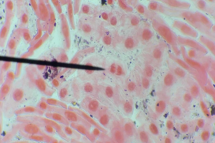
In an article yesterday, I introduced mitotic cell division and the means by which the transition from metaphase to anaphase is controlled, so as to avoid the consequences of mis-segregation of chromosomes, the results of which can be devastating. Today, I shall give an overview of how the wait anaphase signal is generated, as well as how the spindle assembly checkpoint is turned off when proper kinetochore-microtubule attachment has been established.
How Is the Wait Anaphase Signal Generated?
For wild-type yeast cells a spindle defect delays mitotic progression. The molecular components of the spindle assembly checkpoint pathway were first discovered in budding yeast treated with the microtubule-destabilizing drug benomyl. [1,2] The checkpoint components identified in these screens are indispensable in yeast for the spindle checkpoint. These proteins include Mad1, Mad2, Mad3, Bub1, and Bub3. Mad2 mutant cells are not affected by the spindle defect and divide at a normal rate. The spindle defect, however, results in improper segregation of chromosomes and the consequences of this are inevitably lethal.
The wait anaphase signal functions by high-affinity binding of checkpoint component Mad2 to Cdc20, thereby inhibiting the APCCdc20 complex and preventing the ubiquitylation of securin. Progression to anaphase can be blocked by even a single unattached kinetochore, and it is thought that unattached kinetochores catalyze a conformational change in Mad2, thereby allowing it to bind to Cdc20. In support of this hypothesis is the observation that Mad2 is found at some level at unattached kinetochores and appears to rapidly associate and dissociate with the kinetochore.
During prometaphase, a portion of Mad2 is bound to checkpoint component Mad1. Cdc20 is bound and sequestered by a separate portion of Mad2. Mad2 can be bound to only one of those proteins at a time, since it uses the same site to bind Mad1 and Cdc20. Mad2 has been shown to adopt two distinct natively folded states. [3,4,5,6] Mad2 adopts a closed conformation (C-Mad2) when bound to one of its partners, Mad1 or Cdc20. In this state, the carboxy-terminus of Mad2 closes around its binding partner and has been described as a “safety belt.” [7] When Mad2 is not bound to Mad1 or Cdc20, it exists in an open conformation (O-Mad2). In this state, the safety belt is held against the side of Mad2, leading to an inability to efficiently bind Mad1 or Cdc20 until the safety belt has been loosened by a conformational change.
What facilitates this change in Mad2 conformation, thereby generating the wait anaphase signal? One proposed model suggests that the formation of Mad2-Cdc20 complexes at incorrectly attached kinetochores is catalyzed by Mad2-Mad1 complexes. This hypothesis is supported by the fact that C-Mad2 can form a dimer with O-Mad2, initiate a conformational change that loosens the safety belt, and thereby promote its binding to Cdc20. The newly formed C-Mad2-Cdc20 complex is subsequently released from the kinetochore and, in a remarkable positive feedback loop, catalyzes the synthesis of more C-Mad2-Cdc20 complexes by interaction with free O-Mad2 proteins. The C-Mad2-Mad1 complex remains at the kinetochore and repeats the reaction cycle.
A number of other checkpoint components are also involved in inhibition of the APC/C. Inhibitory complexes consisting of Cdc20 and BubR1 (the mammalian homologue of the yeast protein Mad3) and Bub3 are produced by unattached kinetochores. Together, the Mad2-Cdc20 and BubR1-Bub3-Cdc20 complexes suppress the activation of the APC/C. Interestingly, the ubiquitylation by the APCCdc20 of S-phase cyclin A in prometaphase is not blocked by these inhibitory complexes. [8] The explanation for this is unclear.
Turning Off the Spindle Assembly Checkpoint Pathway
Once all of the sister chromatid pairs have been properly bioriented on the mitotic spindle, the APCCdc20 is no longer inhibited, and facilitates the ubiquitin-mediated destruction of securin and M-cyclin. What mechanisms ensure that the spindle assembly checkpoint is turned off following proper biorientation of sister chromatids? Various checkpoint silencing pathways exist. [9] In metazoans, checkpoint components are transported away from kinetochores along microtubules towards the spindle poles in an ATP-dependent manner by cytoplasmic dynein-dynactin motor complexes. [10,11] This process is known as “stripping.” When dynein is inhibited, the removal of Mad1 and Mad2 from the kinetochore is prevented. [12] Indeed, when Mad1 is artificially tethered to correctly attached kinetochores, the onset of anaphase is delayed. [13] Required for recruitment of dynein to kinetochores are the proteins Spindly and RZZ (rough deal, zeste white 10, zwilch). In cases of Spindly motif mutants that are unable to bind dynein, dynein is not recruited to the kinetochore. [14] In such cases, however, the checkpoint is silenced by a second pathway.
A further protein, called p31comet (formerly known as CMT2), has also been associated with checkpoint silencing. [15,16,17] By structural mimicry of Mad2, p31comet is able to bind to Mad2 at the dimerization interface, thereby inhibiting its activity. Another protein that has been shown to be involved in checkpoint silencing by dephosphorylating checkpoint components is protein phosphatase 1 (PP1). [18,19]
The Consequences of Checkpoint Dysfunction
Checkpoint dysfunction can lead to discrepancies in chromosome number (aneuploidy), the consequences of which include tumorigenesis and Down syndrome. [20,21] When spindle checkpoint signaling is reduced in mouse models, a rise in cancer development is observed. [22] The importance and significance of mutations in spindle checkpoint genes are not entirely clear, since spindle checkpoint mutants are relatively infrequent in human tumors, and colon cells exhibiting chromosomal instability appear to possess a fully functional spindle checkpoint. [23] More commonly mutated in such cells are the genes coding for the APC/C. [24,25] Mutations affecting checkpoint genes are not the primary mechanism of checkpoint impairment. A more frequent cause of aneuploidy is alteration in transcriptional regulation resulting in changes in checkpoint protein levels.
An Elegantly Engineered Surveillance System
The spindle assembly checkpoint pathway is an elegantly engineered surveillance system for protecting the cell from the adverse consequences of improper kinetochore-microtubule attachment. Proper attachment of kinetochores to microtubules is monitored by tension-sensing and by detection of attachment of the ends of the microtubules to the kinetochores. Even a single unattached kinetochore is sufficient to trigger the wait anaphase signal, which inhibits activation of the APC/C that drives entry into anaphase. Impairment of the spindle assembly checkpoint pathway can result in aneuploidy, a contributor to cancer and developmental abnormalities such as Down syndrome.
Notes
1. Hoyt, M.A., Totis, L., Roberts, B.T. (1991) S. cerevisiae genes required for cell cycle arrest in response to microtubule function. Cell 66, 507-517.
2. Li, R., Murray, A.W. (1991). Feedback control of mitosis in budding yeast. Cell 66, 519-531.
3. Sironi, L., Mapelli M., Knapp, S., Antoni, A.D., Jeang, K., Musacchio, A. (2002) Crystal structure of the tetrameric Mad1-Mad2 core complex: implications of a ‘safety belt’ binding mechanism for the spindle checkpoint. The EMBO Journal 21(10), 2496-2506.
4. Luo, X., Tang, Z., Xia, G., Wassmann, K., Matsumoto, T., Rizo, J., Yu, H. (2004) The Mad2 spindle checkpoint protein has two distinct natively folded states. Nature Structural and Molecular Biology 11, 338-345.
5. Musacchio, A., Salmon, E.D. (2007) The Spindle Assembly Checkpoint in Space and Time. Nature Reviews Molecular Cell Biology 8, 379-393.
6. Luo, X., Yu, H. (2008) Protein Metamorphosis: The Two-State Behavior of Mad2. Structure 16(11), 1616-1625.
7. Sironi, L., Mapelli M., Knapp, S., Antoni, A.D., Jeang, K., Musacchio, A. (2002) Crystal structure of the tetrameric Mad1-Mad2 core complex: implications of a ‘safety belt’ binding mechanism for the spindle checkpoint. The EMBO Journal 21(10), 2496-2506.
8. Van Zon, W., Wolthuis, R.M. (2010) Cyclin A and Nek2A: APC/C-Cdc20 substrates invisible to the mitotic spindle checkpoint. Biochemical Society Transactions 38, 72-77.
9. Hardwick, K.G., Shah, J.V. (2010) Spindle checkpoint silencing: ensuring rapid and concerted anaphase onset. F1000 Biology Reports 2(55).
10. Howell, B.J., McEwen, B.F., Canman, J.C., Hoffman, D.B., Farrar, E.M., Rieder, C.L., Salmon, E.D. (2001) Cytoplasmic dynein/dynactin drives kinetochore protein transport to the spindle poles and has a role in mitotic spindle checkpoint inactivation. The Journal of Cell Biology 155(7), 1159-1172.
11. Wojcik, E., Basto, R., Serr, M., Scaërou, F., Karess, R., Hays, T. (2001) Kinetochore dynein: its dynamics and role in the transport of the Rough deal checkpoint protein. Nature Cell Biology 3(11), 1001-1007.
12. Howell, B.J., McEwen, B.F., Canman, J.C., Hoffman, D.B., Farrar, E.M., Rieder, C.L., Salmon, E.D. (2001) Cytoplasmic dynein/dynactin drives kinetochore protein transport to the spindle poles and has a role in mitotic spindle checkpoint inactivation. The Journal of Cell Biology 155(7), 1159-1172.
13. Maldano, M., Kapoor, T.M. (2011) Constitutive Mad1 targeting to kinetochores uncouples checkpoint signalling from chromosome biorientation. Nature Cell Biology 13, 475-482.
14. Gassmann, R., Holland, A.J., Varma, D., Wan, X., Civril, F., Cleveland, D.W., Oegema, K., Salmon E.D., Desai, A. Removal of Spindly from microtubule-attached kinetochores controls spindle checkpoint silencing in human cells. Genes and Development 24(9), 957-971.
15. Habu, T., Kim, S.H., Weinstein, J., Matsumoto, T. (2002) Identification of a MAD2-binding protein, CMT2, and its role in mitosis. EMBO Journal 21(23), 6419-6428.
16. Yang, M., Li, B., Tomchick, D.R., Machius, M., Rizo, J., Yu, H., Luo, X. (2007) p31comet blocks Mad2 activation through structural mimicry. Cell 131(4), 744-755.
17. Hagan, R.S., Manak, M.S., Buch, H.K., Meier, M.G., Meraldi, P., Shah, J.V., Sorger, P.K. (2011) p31(comet) acts to ensure timely spindle checkpoint silencing subsequent to kinetochore attachment. Molecular Biology of the Cell 22(22), 4236-4246.
18. Pinsky, B.A., Biggins, S. (2005) The spindle checkpoint: tension versus attachment. Trends in Cell Biology 15(9), 486-493.
19. Vanoosthuyse, V., Hardwick, K.G. (2009) A novel protein phosphatase 1-dependent spindle checkpoint silencing mechanism. Current Biology 19(14), 1176-1181.
20. Shonn, M.A., McCarroll, R., Murray, A.W. (2000) Requirement of the spindle checkpoint for proper chromosome segregation in budding yeast meiosis. Science 289(5477), 300-303.
21. Kops, G.J., Weaver, B.A., Cleveland, D.W. (2005) On the road to cancer: aneuploidy and the mitotic checkpoint. Nature Reviews. Cancer 5(10), 773-785.
22. Rao C.V., Yang Y.M., Swamy M.V., Liu T., Fang Y., Mahmood R., Jhanwar-Uniyal M., Dai W. Colonic tumorigenesis in BubR1+/-ApcMin/+ compound mutant mice is linked to premature separation of sister chromatids and enhanced genomic instability. Proceedings of the National Academy of Sciences 102(12), 4365-4370.
23. Tighe, A., Johnson, V.L., Albertella, M., Taylor, S.S. (2001) Aneuploid colon cancer cells have a robust spindle checkpoint. EMBO Reports 2(71), 609-614.
24. Cahill, D.P., Kinzler, K.W., Vogelstein, B., Lengauer, C. (1999) Genetic instability and Darwinian selection in tumours. Trends in Cell Biology 9(12), M57-60.
25. Rowan, A.J., Lamlum, H., Ilyas, M., Wheeler, J., Straub, J., Papadopoulou, A., Bicknell, D., Bodmer, W.F., Tomlinson, I.P.M. (2000) APC mutations in sporadic colorectal tumors: A mutational “hotspot” and interdependence of the “two hits”. Proceedings of the National Academy of Sciences 97(7), 3352-3357.
This article was originally published at Evolution News & Science Today, on August 27, 2024.
