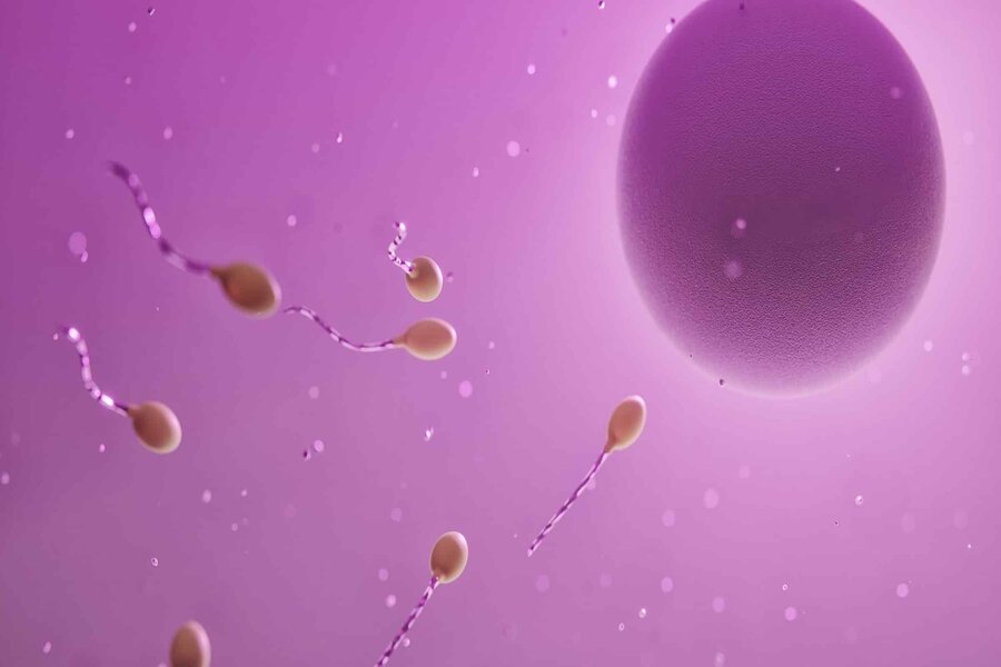
Human reproduction is perhaps the quintessential example of teleology in biology. The process by which a fertilized egg develops into an infant over the space of nine months reveals exquisite engineering and ingenious design. Before this intricate process can even begin, there is a need for a sperm cell to fuse with an ovum — each carrying, in the case of humans, 23 chromosomes. This incredible feat bears the unmistakable hallmarks of conscious intent and foresight. You can watch an animation of this remarkable process here.
In this article, I will focus on the design characteristics of sperm cells. Sperm cells are comprised of three components – the head, the middle piece, and the flagellum – and hundreds of millions of them are carried in the seminal fluid that is released into the cervix through ejaculation during sexual intercourse. With each ejaculation, the male releases between two hundred and five hundred million sperm cells (approximately 100 million per milliliter of semen). Each of these three components, and the seminal fluid, is crucial to the sperm cell’s mission of fusing with an ovum to form a zygote (a fertilized egg). Let us consider each one in turn.
The Head
The head carries densely coiled chromatin fibers, containing the haploid genome — totaling half of the genetic material that will be inherited by the next generation (the other half will come from the mother’s egg cell). The tight packaging of the DNA serves to minimize its volume for transport.
On the tip of the sperm head is a membranous organelle, called the acrosome, that contains various hydrolytic enzymes. When these are secreted, they digest the egg cell membrane, thereby facilitating penetration of the ovum. Without the acrosome, the sperm cell will be unable to penetrate the egg cell membrane to fertilize the ovum. According to a review paper published in Frontiers in Cell and Developmental Biology [1]:
Any structural or functional acrosomal abnormality could impair sperm fusion, and ultimately result in infertility. Moreover, studies have shown that intra-cytoplasmic insemination with sperm containing acrosomal abnormalities did not lead to successful fertilization, even in the absence of fertilization barriers, because the oocyte was unable to be efficiently activated…Thus, the acrosome is indispensable for fertilization.
When a sperm reaches the vicinity of the egg, it undergoes a series of molecular interactions with the zona pellucida, which is a specialized extracellular matrix surrounding the egg. Specific receptors on the sperm’s plasma membrane, such as spermadhesins or integrins, recognize and bind to corresponding ligands on the zona pellucida. This binding triggers the activation of signaling pathways in the sperm. Binding of the sperm receptors to the zona pellucida ligands leads to an influx of calcium ions (Ca2+) into the sperm cell. This calcium influx is typically mediated by ion channels or receptors on the sperm’s plasma membrane, which are activated upon ligand-receptor binding. The increase in intracellular calcium levels initiates a signaling cascade within the sperm cell. Calcium ions act as second messengers and trigger the activation of various downstream signaling molecules and enzymes, including protein kinases. As a result of the calcium-mediated signaling cascade, the acrosome undergoes exocytosis. The membrane surrounding the acrosome fuses with the sperm’s plasma membrane, causing the release of the acrosomal contents, including enzymes such as hyaluronidase and acrosin. The enzymes released from the acrosome help degrade the glycoprotein matrix of the zona pellucida, allowing the sperm to penetrate and reach the egg’s plasma membrane. The acrosomal contents aid in the breakdown of the protective layers surrounding the egg, facilitating the fusion of the sperm and egg membranes.
The formation of the acrosome itself is divided into four stages. The first stage, the “Golgi phase,” is dependent upon the Golgi apparatus, which produces and packages the proteins and enzymes needed for acrosome formation. These proteins are then transported into the developing acrosome vesicle. In the second phase, the “cap phase,” the Golgi-derived vesicle (known as the proacrosomal vesicle) fuses with the anterior portion of the nucleus, forming a cap-like structure over the nucleus. The fusion of the vesicle with the nucleus is mediated by membrane trafficking processes. The proacrosomal vesicle contains enzymes, glycoproteins, and other components that are essential for acrosome maturation. In the third phase, the “acrosome phase,” the cap-like structure undergoes a series of structural changes, leading to the formation of the acrosome. The proacrosomal vesicle flattens and elongates, spreading over the anterior region of the nucleus. The Golgi-derived enzymes modify the proteins present in the proacrosomal vesicle, converting them into their active forms. The acrosomal membrane also undergoes changes, becoming specialized for the acrosome’s functions. In the final phase, the “maturation phase,” the acrosome undergoes further modifications and maturation. Enzymes within the acrosome become fully activated and the acrosomal matrix undergoes changes, becoming more condensed. The acrosomal granule, which is the central region of the acrosome, becomes highly electron-dense due to the accumulation of enzymes and proteins. The mature acrosome is now ready for its role in fertilization. For a more detailed description of this incredible process, I refer readers to a review paper on the “Mechanism of Acrosome Biogenesis in Mammals.” [2]
The Middle Piece
The middle piece consists of a central filamentous core, around which are many strategically placed mitochondria that synthesize the energy molecule adenosine triphosphate (ATP). The complexity and design of energy generation within the mitochondria — including the processes of glycolysis, the citric acid (or, Krebs) cycle, the electron transport chain, and oxidative phosphorylation — could be its own series of articles, but this is a topic for another day. For a good introduction to the phenomenal processes within the mitochondria, here are three animations from Harvard University that bring this fascinating organelle to life:
The ATP generated by the mitochondria energizes the power strokes of the flagellum, driving its journey through the female cervix, uterus, and uterine tubes. As such, the middle piece of the sperm cell is absolutely essential to its function of swimming through the female uterus and fallopian tube to fertilize her egg. Without the middle piece and its mitochondria, the sperm cells are completely immobile.
The Flagellum
Unlike a bacterial flagellum (which rotates like a motor), a sperm flagellum beats with a whip-like motion to produce motility. How does the flagellum work? In 2018, Jianfeng Lin and Daniela Nicastro elucidated the mechanism of flagellar motility. [3] Their data indicated that “bending was generated by the asymmetric distribution of dynein activity on opposite sides of the flagellum” [4] (dyneins are ATP-powered molecular motors that “walk” along microtubules towards their minus end). Their results also revealed that alternating flagellar bending occurs due to “a ‘switch-inhibition’ mechanism in which force imbalance is generated by inhibiting…dyneins on alternating sides of the flagellum.” [5] In other words, regulatory signals lead to the inhibition of dynein motors on one side of the flagellum. Meanwhile, on the other side, the dyneins walk along the microtubules. The flagellum bends in one direction due to molecular linkers that resist this sliding. The flagellar bending alternates by repeatedly switching the side of dynein inhibition. Look here for an animation showing how this is thought to work.
It goes without saying that, without the flagellum, the sperm cell is completely immotile and has no chance of fertilizing the egg.
Thus far, we have considered the irreducible complexity of the components of a sperm cell. We shall now consider the design features of the seminal fluid and the process of sperm capacitation that takes place within the female reproductive tract.
The Seminal Fluid
As I mentioned previously, between two hundred and five hundred million sperm, surrounded by seminal fluid, are released with each ejaculation. Such huge numbers are necessary in order to have a significant chance of fertilizing the egg, since many hazards confront the sperm cells as they swim through the uterus and uterine tubes. Following ejaculation, millions of the released sperm cells will either flow out of the vagina, or else die in its acidic environment. Sperm cells also need to pass through the cervix and opening into the uterus, which requires passage through the cervical mucus. Though the mucus is thinned to a waterier consistency during the fertile window, making it more hospitable to sperm, millions of sperm cells will nonetheless die attempting to make it through the mucus. Furthermore, the female reproductive tract has immune defenses that protect against pathogens. These defenses can also target and destroy foreign cells like sperm. Antibodies may recognize sperm as foreign invaders and lead to their inactivation or elimination. There are also tiny cilia in the fallopian tube that propel the egg towards the uterus. Some of the remaining sperm will become trapped in the cilia and die. Only a small handful of the original sperm cells will make it as far as the egg. Thus, it is necessary that hundreds of millions of sperm cells are released in order to have a reasonable chance of the egg cell being fertilized.
Seminal fluid also provides essential nutrients to support the survival and motility of the sperm. These include fructose — which serves as a source of energy for the sperm, fueling the mitochondrial production of ATP — as well as other sugars, amino acids, and enzymes. If the seminal fluid did not contain fructose, to power the mitochondria, this would have drastic implications for sperm cell motility and viability.
The seminal fluid is also alkaline. This is important because the vagina has an acidic pH, produced by the normal flora (bacterial populations) of the vagina. This environment would be unfavorable to sperm cells. But the alkalinity of the seminal fluid helps to neutralize the vagina’s acidic pH, assisting the survival of the sperm.
Following ejaculation, the seminal fluid initially coagulates to form a gel-like consistency. This coagulation helps to keep the semen in the vagina and cervix, preventing it from immediately leaking out and thereby greatly increasing the odds of a successful fertilization. This occurs upon exposure to the air or the alkaline environment of the female reproductive tract, activating clotting factors present in the seminal fluid, including tissue transglutaminase. The transglutaminase converts semenogelin (a major protein in seminal fluid secreted by the seminal vesicles) into a sticky protein called fibrin. Fibrin forms a network-like structure that entraps sperm and other components of the semen.
If the semen remained in this state, the sperm would be permanently immobile and unable to fertilize the egg. Over time, however, the coagulated semen liquefies due to enzymes present in the fluid that slowly break down the fibrin network, allowing the sperm to move more freely. Anamthathmakula and Winuthayanon note that “The liquefaction process is crucial for the sperm to gain their motility and successful transport to the fertilization site in Fallopian tubes (or oviducts in animals). Hyperviscous semen or failure in liquefaction is one of the causes of male infertility.” [6] In fact, targeting these serine proteases has been suggested as a target for novel non-hormonal contraceptives. [7]
From an evolutionary perspective, it is difficult to envision a scenario where semen coagulation evolved, without simultaneously having a mechanism for liquefaction. This is a prime example of a non-adaptive intermediate that is prohibitive to evolution by natural selection.
Sperm Capacitation
In order for a sperm cell to fertilize an egg, it has to undergo capacitation. This takes place in the female reproductive tract. The process of capacitation involves a series of biochemical and physiological changes that prepare the sperm for successful interaction with the egg and is crucial in order for the sperm cell to acquire the ability to fertilize.
When sperm are initially ejaculated, they possess certain molecules and proteins on their surface that inhibit their ability to fertilize an egg. During capacitation, these surface molecules, such as cholesterol and glycoproteins, are removed or modified, allowing the sperm to become more receptive to the egg. As capacitation progresses, the motility pattern of sperm also changes. They undergo hyperactivation, which is characterized by increased amplitude and asymmetrical beating of the tail. Hyperactivated sperm exhibit vigorous movements, which help them to navigate through the female reproductive tract and reach the egg. Capacitation also involves changes in the composition and fluidity of the sperm cell membrane. These changes allow the sperm to better interact with the egg’s zona pellucida. The acrosome becomes primed for the acrosome reaction, which releases these enzymes to allow penetration of the egg membrane.
Capacitation is associated with an increase in calcium ion influx into the sperm. Calcium plays a crucial role in various intracellular signaling processes that are necessary for sperm function and fertilization. For a much more detailed treatment of what is known about the mechanisms of sperm capacitation, there are good reviews of this subject, to which I direct readers. [8,9]
Conclusion
In summary, various features of the head, middle piece, and flagellum, together with the properties of the seminal fluid, are critical to the sperm cell’s function of reaching and fertilizing an egg. If any one of these parts is not present or fails to function properly, the sperm cell is rendered completely impotent, and reproduction cannot occur. The phenomenon of human reproduction points to a cause with foresight — one that can visualize a foreordained outcome and bring together everything needed to realize that end goal. There is no cause in the universe that is known to have such a capacity of foresight other than intelligent design.
Footnotes
1. Khawar MB, Gao H, Li W. Mechanism of Acrosome Biogenesis in Mammals. Front Cell Dev Biol. 2019 Sep 18;7:195.
2. Ibid.
3. Lin J, Nicastro D. Asymmetric distribution and spatial switching of dynein activity generates ciliary motility. Science. 2018 Apr 27;360(6387):eaar1968.
4. Ibid.
5. Ibid.
6. Anamthathmakula P, Winuthayanon W. Mechanism of semen liquefaction and its potential for a novel non-hormonal contraception†. Biol Reprod. 2020 Aug 4;103(2):411-426.
7. Ibid.
8. Puga Molina LC, Luque GM, Balestrini PA, Marín-Briggiler CI, Romarowski A, Buffone MG. Molecular Basis of Human Sperm Capacitation. Front Cell Dev Biol. 2018 Jul 27;6:72.
9. Stival C, Puga Molina Ldel C, Paudel B, Buffone MG, Visconti PE, Krapf D. Sperm Capacitation and Acrosome Reaction in Mammalian Sperm. Adv Anat Embryol Cell Biol. 2016;220:93-106.
Note: This essay has been adapted from a two-part series of articles originally published at Evolution News & Views (here and here), on June 30th and July 3rd 2023.

2 thoughts on “On the Irreducible Complexity of Sperm Cells”
Pingback: For Males, an Engineering Marvel that Originates in the Brain - Jonathan McLatchie | Writer, Speaker, Scholar
Pingback: How NOT to Argue Against Irreducible Complexity - Jonathan McLatchie | Writer, Speaker, Scholar
Comments are closed.